Gliomas in dogs
11 June 2026
In 2025, Dr Andy Yale from the RVC received BSAVA PetSavers funding to support a student project investigating immune checkpoint proteins and novel immunotherapy targets in canine glioma. Student Sunita Garg describes her work:
Gliomas in dogs
Gliomas are the second most common brain tumours in adult dogs, and comprise two main subtypes: oligodendrogliomas (approximately 70% of all gliomas) and astrocytomas (20% of all gliomas)(1). Older male dogs, particularly brachycephalic breeds such as Boston Terriers and French Bulldogs, are more commonly affected (2). Currently, dogs with glioma have a poor prognosis due to a lack of efficacious treatments, as well as difficulty in diagnosing the tumours at earlier stages. Surgery is often challenging due to the tumour intra-axial location and diffuse margins. Radiation therapy can improve outcome in some dogs, although prognosis is still poor and the treatment is invasive and costly. Chemotherapy is largely ineffective with short median survival times of only 1–3 months (1,3).
Immunotherapy is an increasingly popular and effective cancer treatment, often aimed at overcoming immune system suppression by tumour cells. This is a common evasion tactic employed by a range of tumours, usually through the upregulation of immune checkpoint proteins such as CTLA-4 and PD-L1. Certain immunotherapy treatments aim to inhibit these proteins, and have been successful in both humans and canines in treating various types of cancer (4).
Our aim was to determine whether two potential immunotherapy targets, PD-L1 and CTLA-4, are upregulated in canine gliomas. These proteins are known to be upregulated in a proportion of human gliomas and other types of canine tumours. Therapeutic monoclonal antibodies have also been developed against both targets in dogs, meaning clinical trials targeting PD-L1 and CLTA-4 could be considered for canine glioma in the near future, should their expression be confirmed in this tumour. This was a pilot study aimed at validating the PD-L1 and CTLA-4 antibodies in canine tissue, prior to assessing their expression in a small number of canine glioma and normal brain samples.
Our findings
Using a sample of nine canine glioma tissues and four normal brain tissues from the RVC Companion Animal Brain Bank and RVC Pathology Department, we produced haemoxylin and eosin (H&E) slides for each tissue. Gliomas were then categorised into histologic subtypes and graded, with most tumours being of a high grade. After this, we performed CTLA-4 and PD-L1 immunohistochemistry (IHC) with commercial antibodies on all glioma and normal brain samples. Since the antibodies were developed primarily for use in human and murine tissue, we also used canine tonsil and lymph node tissue as a positive control, to determine the specificity of the antibody binding in canine tissue.
Alongside this, to further confirm antibody specificity in canine tissue, we performed western blot analysis. Using kidney tissue as a negative control, and lymph node and tonsil tissues as a positive control, we were able to validate the use of our antibodies in canine tissues (Figure 1), with the exception of CTLA-4 where we struggled to optimise the western blot.
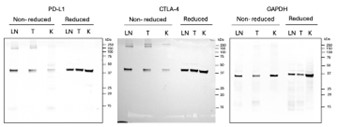
Fig 1. Western blot membrane showing staining with PD-L1 (expected band size is around 40kDa), CTLA-4 (expected band size is 25-30 kDa), and GAPDH (expected band size is 36kDa). No bands are present for CTLA-4 (visible bands are from previous staining with PD-L1).
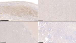
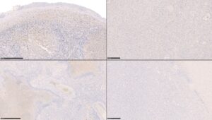
Results from the IHC are presented in Figures 2 and 3. Positive labelling for PD-L1 and CTLA-4 was observed in positive control tissue. We did not identify any PD-L1 or CTLA-4 labelling in canine glioma tissue. Although these checkpoint proteins are only expressed in a small proportion of human glioma, our study results suggest they are expressed in an even lower proportion of canine glioma. While this may signify that these proteins are not good targets for immunotherapy in dogs, they must be explored in a larger population before drawing this conclusion. Other protein targets should also be explored as future immunotherapy targets, such as epidermal growth factor receptor.
Challenges we faced
During the course of this project, one of the biggest challenges we faced was establishing that the antibodies used were successful in identifying the correct proteins in canine tissues. For this, alongside performing IHC in control tissue, we also performed western blot analysis. Having done very little wet lab work previously, I personally found this a complicated process. However, with the help of the RVC team, we were able to produce successful blots except for CTLA-4. We suspect this is due to a low expression of CTLA-4 in non-reactive lymphoid tissue and plan to further optimise the CTLA-4 western blot by repeating the analysis in reactive lymph node tissue or another tumour type where CTLA-4 is often upregulated (such as oral melanoma). Another challenge was interpreting histology and IHC slides of gliomas, as I have done little pathology work previously. Overcoming these problems was highly rewarding and has better prepared me for future research and clinical work following graduation.
How might these results impact general practitioners?
Whilst immunotherapy is already a treatment option for some canine cancers and non-neoplastic conditions, being able to develop immunotherapy for gliomas could provide an incredibly valuable treatment option for vets and owners, as current treatment options have limited success. It would not be unreasonable to suggest that immunotherapy could be administered in general practice.
What this experience meant to me
This research placement enabled me to develop my skills in a range of topics, both research-based and clinical. This included writing papers, interpreting histological slides, and performing wet lab experiments. Working alongside the clinicians at the RVC has given me a clearer insight into the role of veterinarians in research, something I wish to pursue following my graduation, and has been invaluable in my development as a veterinarian.
I am thankful to BSAVA PetSavers for funding this project. It has been invaluable to me personally, and has also helped improve our current knowledge of canine gliomas and their treatment. I would highly recommend any students thinking of doing a research project, either as part of their EMS or otherwise, to apply for a student research project grant.
Author biography

References
- Miller AD, Miller CR, Rossmeisl JH. Canine Primary Intracranial Cancer: A Clinicopathologic and Comparative Review of Glioma, Meningioma, and Choroid Plexus Tumors. Front Oncol [Internet]. 2019 Nov 8 [cited 2025 July 14];9. Available from: https://www.frontiersin.org/journals/oncology/articles/10.3389/fonc.2019.01151/full
- José‐López R, Gutierrez‐Quintana R, De La Fuente C, Manzanilla EG, Suñol A, Pi Castro D, et al. Clinical features, diagnosis, and survival analysis of dogs with glioma. J Vet Intern Med. 2021 July;35(4):1902–17.
- José-López R. Chemotherapy for the treatment of intracranial glioma in dogs. Front Vet Sci. 2023 Oct 31;10:1273122.
- Shiravand Y, Khodadadi F, Kashani SMA, Hosseini-Fard SR, Hosseini S, Sadeghirad H, et al. Immune Checkpoint Inhibitors in Cancer Therapy. Curr Oncol. 2022 Apr 24;29(5):3044–60.
Supporting more BSAVA PetSavers research
Help us fund more clinical veterinary research into companion animal diseases by donating today at www.bsava.com/petsavers/donate or by scanning the QR code: